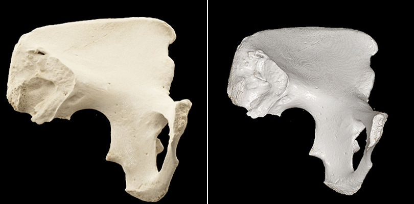
Researchers from Thailand used cinematic rendering to enhance the visualization of pelvic bone CT scans, enabling radiologists and osteologists to identify key features for age estimation without invasive preparation, according to an article recently published in Forensic Science International.
Cinematic volume rendering boosted the accuracy of physicians in identifying pelvic bone features for age estimation to more than 91% overall, compared with the 81% accuracy of evaluations with traditional volume rendering reported in prior studies, first author Dr. Nuttaya Pattamapaspong, an associate professor of radiology at Chiang Mai University, told AuntMinnie.com (Forensic Sci Int, January 2019, Vol. 294, pp. 48-56).
“This volume rendering technique helps us more clearly understand complex structures and their 3D relationship,” he said. “I believe this has the potential to improve the quality of surgical planning, patient consultation, and medical education.”
Bone evaluation gets new look
The photorealistic display of cinematically rendered CT scans has become increasingly more relevant for the visualization of intricate anatomy. Several groups have highlighted the technique’s usefulness in the clinical evaluation of ovarian cancer, acute aortic injury, and kidney aneurysms.
What’s more, a study out of Switzerland revealed that radiologists and orthopedic surgeons favored the realistic depiction afforded by cinematic rendering over traditional volume rendering for the assessment of ankle fractures.
In the present study, Pattamapaspong and colleagues explored the possibility of applying cinematic rendering to the inspection of bone morphology, with the particular goal of improving age estimation based on bone features.
The researchers began by acquiring CT scans of 35 dry pelvic bones and then converting them into 3D reconstructions with cinematic rendering software (syngo.via VB20, Siemens Healthineers). The average age of the bones at death was 48.5 years, and 40% of the bones came from women.

Two radiologists and a forensic osteologist independently examined the cinematically rendered CT scans. They were able to detect key age features with a near-perfect level of accuracy in the pubic symphysis (the midline of the pelvic bone) but only moderate accuracy in the auricular surface (upper anterior region), compared with the gold standard of dry bone interpretation by an anatomist.
For detecting features near the pubic symphysis, the clinicians slightly overstaged or understaged patterns for three of the pelvic bones but otherwise identified all other features with almost 100% accuracy. Their characterization was also excellent for the uppermost regions of the auricular surface at 91.4% accuracy, but accuracy fell to approximately 70% for lateral regions of the auricular surface and to about 50% for porosity (diameter of the holes in the pelvic bone).
Intra- and interobserver agreement was nearly perfect for all features around the pubic symphysis and substantial for the top portion of the auricular surface, but agreement was only moderate or fair for porosity.
Further optimization required
Overall, the accuracy of detecting key features on cinematically rendered CT scans was more than 10 percentage points greater than the detection accuracy reported by Telmon et al in a previous study that relied on traditional volume rendering techniques for visualization.
The high percentage of correct interpretations and the high level of agreement between cinematically rendered CT scans and the gold standard suggests that the new technique is effective for displaying age-related changes near the pubic symphysis of pelvic bones, the authors noted. However, the technique’s application does not meet the standard for evaluating the auricular surface of the bones.
“3D CT scans are not yet able to replace manual examination of skeletons, but do offer additional advantages to the examination of specimens, such as improving the visualization of poorly preserved or obscured areas and reducing potential problems from invasive analysis,” Pattamapaspong said.
The radiation dose and very thin slice sections used during CT acquisition of the pelvic bone allowed for its high-resolution display, he added. These parameters may need to be adjusted when performing CT exams of actual patients, and, therefore, appropriate use of the technique may lead to different results in a clinical setting.
“Our study gives an example of the anticipated level of efficacy and the current limitations of pelvic CT bone examination using cinematic rendering technology,” he said. “With emerging virtual reality technologies, application of new tools in forensic medicine and anthropology will require adjustments, technical optimization, and validation.”
Copyright © 2019 AuntMinnie.com. Source: Cinematic rendering enhances pelvic CT bone evaluation


